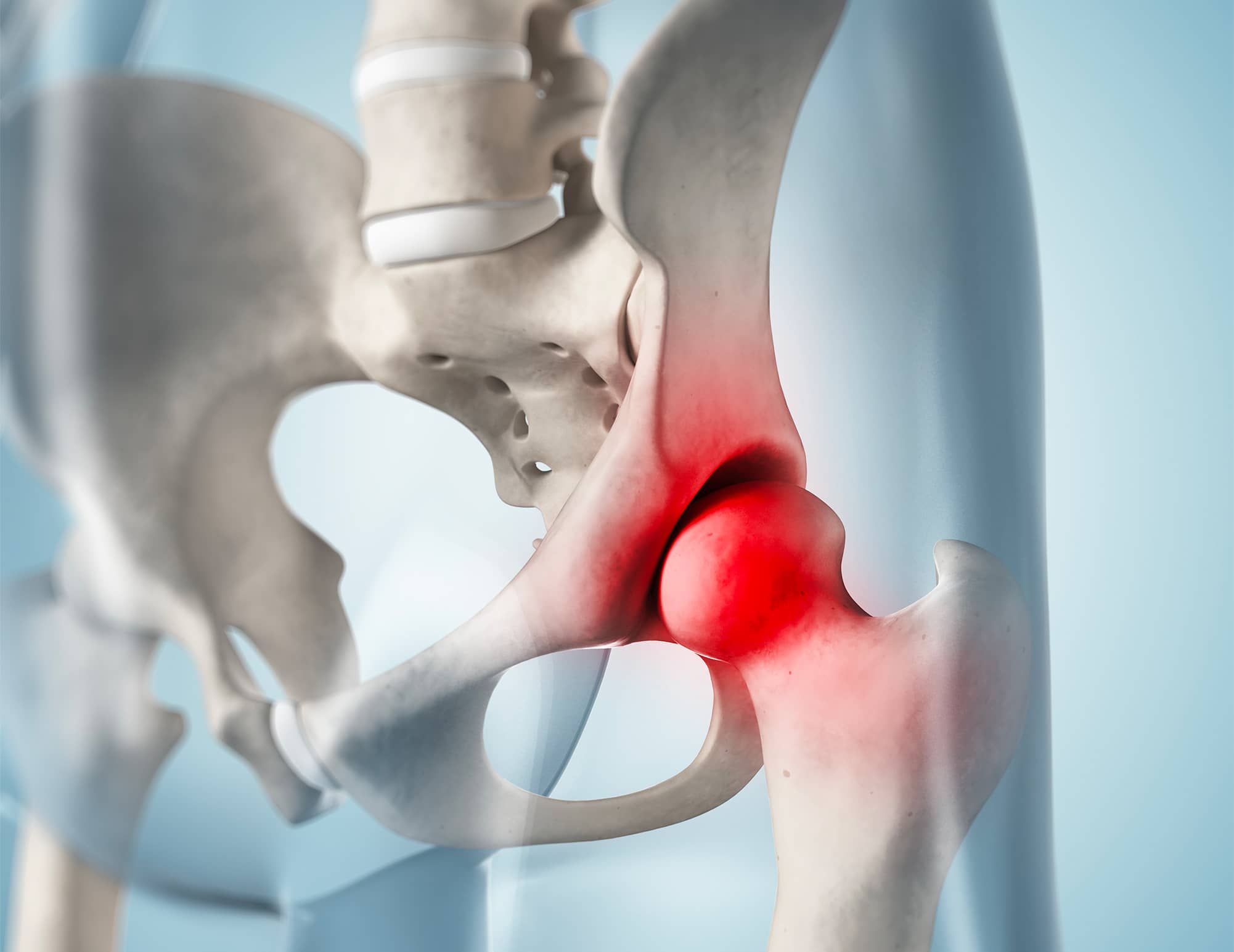
Synovial Chondromatosis
Synovial Chondromatosis of the hip is a rare condition, often considered in young male patients presenting with hip pain and normal radiographs. It is a benign pathology characterized by the formation of cartilaginous nodules (chondromas) within the joint, which may subsequently ossify into osteochondromas. Hip arthroscopy is used to confirm the diagnosis and to perform minimally invasive treatment.
Diagnosis of Synovial Chondromatosis is often challenging due to the depth of the joint. Primary Synovial Chondromatosis is a disease of the synovial membrane with an unknown origin. This pathological synovial membrane produces cartilaginous nodules called chondromas, which can become pedunculated and then detach, forming free foreign bodies in the joint cavity. The chondromas can grow and sometimes ossify into osteochondromas, leading to osteochondromatosis. Secondary Chondromatosis, on the other hand, is a different disease associated with osteoarthritis, which releases cartilaginous debris.

Diagnosis
Synovial Chondromatosis progresses slowly with late clinical signs. The pain is intermittent and movement-related (mechanical pain). Internal disturbances such as painful locking or joint snagging are typical. Advanced chondromatosis can lead to joint stiffness.
When the chondromas are calcified or ossified, standard radiography easily establishes the diagnosis in the stage of osteochondromatosis. However, in cases of chondromatosis where the chondromas are not ossified, radiographs are often normal.
Arthro-CT scanning is the best diagnostic tool for chondromatosis. It allows visualization of chondromas, osteochondromas, synovial membrane thickening, and cartilage alterations that determine the disease’s prognosis. Arthro-MRI provides the same information as Arthro-CT.
Despite Arthro-CT, the diagnosis of chondromatosis can sometimes remain doubtful. In such cases, hip arthroscopy is useful. It can confirm the diagnosis and sometimes correct a mistaken one. Discovering chondromatosis can be surprising when exploring a “mysterious” painful hip.
Clinically, the condition evolves with recurrent flare-ups. Eventually, it progresses to secondary osteoarthritis (coxarthrosis). Long-term monitoring of synovial chondromatosis is justified to detect recurrence, secondary coxarthrosis, and in rare cases, a malignant transformation of the disease (sarcoma).
Treatment
Acetabular chondropathies are common, often associated with hip impingement or dysplasia. If medical treatment is ineffective, hip arthroscopy is considered to correct the cause of impingement, repair the labrum lesion, and resect unstable cartilage lesions. Subchondral perforations are also performed during arthroscopy if the subchondral bone is exposed. In cases of severe dysplasia, a periacetabular osteotomy or prosthesis implantation is discussed.
Femoral chondropathies are rarer and can be associated with osteochondritis of the femoral head, sometimes in a traumatic context. Arthroscopic treatment allows for the resection of unstable cartilage lesions. Osteochondral grafts are exceptionally proposed.
When cartilage lesions are severe and associated with advanced osteoarthritis, treatment relies on prosthesis implantation. In young and athletic individuals, a custom implant, implanted via the anterior approach, is preferred.